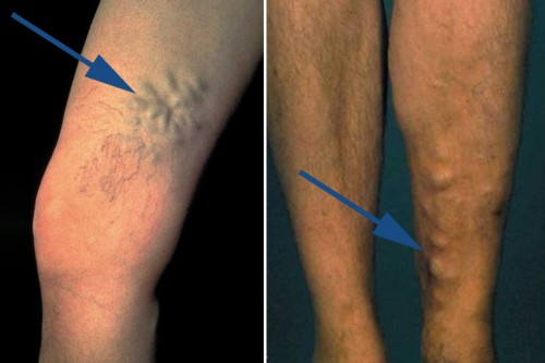
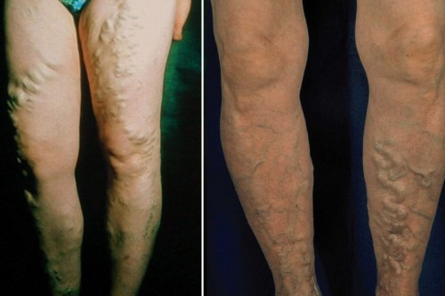
About 30% of all women and men suffer from large varicose veins
Here you find more information about the different types of varicose veins, their symptoms, and the complications

Definition & occurrence of varicose veins
Varicose veins are pathologically dilated, superficial veins, often appearing as twisted or nodular structures in the legs. In technical terms, they are also called varicosities. They lie in or closely beneath the skin, so they can usually be seen clearly. Varicose veins affect only the veins of the superficial vein system.
The smallest varicose veins are the spider veins and the largest are varicose veins of the side branch or trunk veins.
When varicose veins are present, the condition is referred to as varicose disease or varicosis.
The condition is chronic and progressive, which means that new varicose veins may develop with time despite treatment. The predisposition to varicose veins, that is to say, the underlying connective tissue weakness, cannot be cured. And there is no way of overcoming some of the varicose vein risk factors, such as advancing age. Therefore, there is no therapeutic approach that can provide a permanent cure and new varicose veins can develop again.
Primary varicose disease
Varicose disease can be divided into primary and secondary forms. Most cases are primary disease, which means that varicose veins develop because of a hereditary weakness in the connective tissues and vein walls. These are the typical varicose veins that are dealt with on this website.
Secondary varicose veins are the result of another vein disease; for example, when a deep vein thrombosis causes varicose veins after the thrombosis event. The prior disease is the cause of these (secondary) varicose veins and it also needs to be treated.
Types of varicose veins
Varicose veins of the legs are divided into spider veins and reticular varicose veins (small varicose veins), varicose veins of the perforating, side branch, and trunk veins.
Small varicose veins
Spider veins are dilated veins measuring up to 1 mm, while reticular veins have diameters of 1-3 mm. Both lie in the skin, the spider veins somewhat more superficially (nearer the surface) than the reticular veins.
The small varicose veins have already been described in detail in the section on spider veins.
Perforating varicose veins
Perforating varicose veins arise from poorly functioning connecting veins between the superficial and deep vein systems. Each leg has about 150 of these perforating veins (perforators). They allow blood to flow from the superficial veins into the deep veins. The perforating veins also contain valves to prevent the backwards flow of blood. If these valves are leaky, the blood flows in the wrong direction from the deep to the superficial veins, causing varicose veins to develop over time.
Perforating veins that are frequently affected are Dodd’s and Hunter’s perforators found on the inside of the legs above the knee and the three Cockett’s perforators above the inside of the ankle.
Varicose veins of perforators are often characterised by a balloon-like dilatation, the blow-out phenomenon. Increased pressure in the vein due to the reversed blood flow causes the vein to dilate like a balloon, bulging out the overlying skin. The vein is then particularly close to the surface and bleeds easily after only minor injuries.
Varicose veins of the side branch or tributary veins
Varicose veins of the side branch or tributary veins develop when valves in these veins no longer close tightly and the veins dilate because of the backflow of blood. The disease is also known as side branch varicosis and is often associated with or caused by trunk varicose veins. Side branch varicose veins may, however, also occur in isolation.
These varicose veins are usually very tortuous and clearly visible as they make considerable bulges in the skin.
Trunk varicose veins
Trunk varicose veins arise when the valves in the trunk veins (the great saphenous vein/vena saphena magna and the small saphenous vein/vena saphena parva) no longer close tightly and are affected by varicose disease. On examination of the legs while the patient is standing up, doctors are able to distinguish different stages of disease (Hach stages), depending on which valves are affected and how far the blood flows back down the leg. Considering the great saphenous vein, it could be that only the valve in the groin is leaking or the blood backflow may reach further down the leg: to the knee, to below the knee or even as far as the ankle. The picture is similar for the small saphenous vein – with either only the first (terminal) valve being affected or with the backflow extending down the calf or even as far as the ankle.
The trunk veins usually lie so deep that these varicose veins cannot be seen with the naked eye, but they often occur together with side branch varicose veins that are clearly visible.
The precise anatomical sites of the different types of varicose veins are summarised in the following diagram:
Occurrence of varicose veins
Germany
Varicose veins are one of the most common diseases in the German population. The incidence of the disease increases with age, so that almost every elderly person has some form of changes in the leg veins.
About 60% of adults in Germany have spider veins and/or small varicose veins without larger varicose veins, while about 31% of adult Germans have large varicose veins requiring treatment.
Women are somewhat more often affected than men.
In Germany, about 34% of women and 28% of men have pronounced varicose veins, so the gender difference is not actually that big. In some countries, however, men seem to be much less commonly affected than women. The reason why German men are particularly frequently affected by varicose veins remains unclear.
Worldwide occurrence
In Germany, about 31% of adults suffer from large varicose veins, whereby about 4% already have skin changes and/or venous leg ulcers (Rabe, Phlebologie 2003;32:1). An international study (Rabe, International Angiology 2012;31:105) investigated the prevalence of vein disease worldwide. The following table shows the results.
| Prevalence of varicose veins worldwide | |||
| Geographical region | Persons with “only” spider and reticular veins | Persons with large varicose veins | Persons with skin changes/venous ulcers |
| Germany | 60% | 31% | 4% |
| Western Europe | 43% | 34% | 7% |
| Central + Eastern Europe | 38% | 49% | 12% |
| Latin America | 42% | 46% | 13% |
| Middle East | 54% | 31% | 7% |
| Far East | 45% | 42% | 7% |
It can be seen at a glance that the concept of varicose veins as a lifestyle disease of westernised countries is simply not true: such veins also occur in developing countries and Asia. Varicose disease is also common in the Middle East and the Far East. Compared with Germany, other regions are even more widely affected, particularly regarding the serious complications of skin changes and venous leg ulcers. Germany clearly has the lowest incidence of such complications, while rates are particularly high in Latin America and Central and Eastern Europe. This finding may be due to the fact that varicose disease is taken more seriously in Germany than elsewhere and early treatment prevents most serious complications.
In summary, it can be said that varicose disease represents a real worldwide health problem that needs to be taken seriously. Although regional differences do exist, the number of people affected by varicose veins is relatively constant throughout the world, a fact that makes it a common global disease.


