Optimal diagnostic investigation of the veins is the basis for successful treatment
These investigations are non-invasive and painless

Examination for the diagnosis of spider veins and varicose veins
Examinations needed to make the diagnosis
The vein specialist will make a thorough examination. These days, the investigations needed for the diagnosis of varicose veins are non-invasive and painless.
First of all, the doctor will take your medical history, for example, how long you have had the varicose veins and what symptoms they cause.
To determine your risk factors, you will also be asked whether any other members of your family have varicose veins and whether there are any aspects of your lifestyle that could be improved. If you have already had treatment, the doctor will enquire about any previous therapeutic measures.
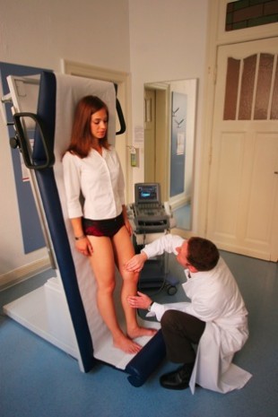
After the medical history comes the physical examination. The doctor will carefully examine and palpate your legs, while you are standing up, thereby looking for any visible varicose veins, swelling, and skin changes.
Any warmth of the skin or tenderness over a particular area may indicate inflammation of the underlying vein.
Ultrasound examination of the leg veins is nowadays a routine method of diagnosis.
Optimal diagnostic investigation of the veins with ultrasound
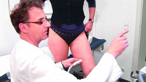
Doppler or duplex ultrasound scanning is now a routine procedure in the diagnosis of varicose veins. It is completely painless, carries no risks, and does not involve any exposure to radiation. With the aid of this technique, the affected areas of the veins can be visualised and the extent of the disease accurately determined. This is necessary for drawing up an optimal treatment plan. Depending on the extent of the problem, the examination lasts 5-20 minutes.
The ultrasound examination
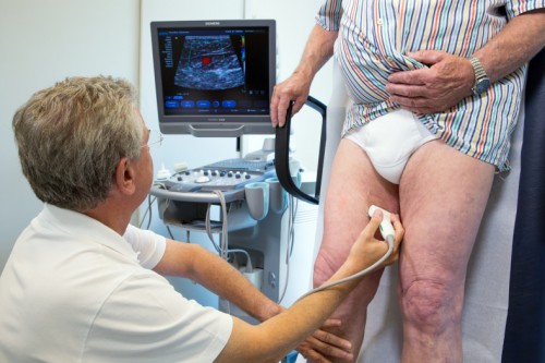
This examination provides images of various parts of the body by means of ultrasound waves. First of all, contact gel is applied to the relevant area of skin, as any air trapped between the ultrasound probe and the skin would interfere with the examination. The probe is held over the vein in question and sends out ultrasound waves, which pass through the skin into the tissues. These sound waves are absorbed to varying extents by the different layers of tissue and are reflected back accordingly. The probe picks up the reflected waves, thus acting as both sound wave transmitter and receiver.
A grey scale display system is used for the returning waves and the image appears on the screen in black and white. This is known as B-mode imaging and is currently the ultrasound examination most widely used in medicine.
Doppler ultrasonography
Doppler ultrasound is used to measure blood flow in the heart and vessels. If the sound waves hit blood cells in the vessels, some of the waves are reflected back with a different frequency. This change in frequency depends on the movement of the blood cells and thus allows their flow direction and velocity to be measured. The results can be presented in various ways, for example, as an audible signal or as a flow curve on the display screen.
Duplex ultrasonography
Duplex ultrasound is a combination of routine ultrasound imaging of the tissues (B-mode ultrasound, black/white imaging) and Doppler ultrasound (measurement of blood flow).
Duplex ultrasound is the most up-to-date technology for diagnosing vein disease, although ideally it is not only used for diagnosis, but also to monitor disease progression and the results of treatment after sclerotherapy or surgery. Duplex ultrasound can be used to look at superficial veins, deep veins, arteries, and the tissues surrounding the vessels on the display screen. It is also possible to show the rate and direction of the blood flow within the vessels in different colours. Blood flowing towards the probe appears red, away from the probe is shown in blue.
The examination provides important information on thrombosis, disorders of the valves, any possible backflow towards the feet, the extent of disease, and the prognosis of the vein problems.


